Thrombus Aspiration Used Alone to Treat ST Elevation Myocardial Infarction Patients with Chronic Atrial Fibrillation
Article information
Trans Abstract
The safety and effectiveness of thrombus aspiration alone without additional ballooning or stent implantation in ST-segment elevation myocardial infarction (STEMI) patients is limited. Recently, we experienced a case of a STEMI patient with chronic atrial fibrillation who was successfully treated with thrombus aspiration alone.
Introduction
ST-segment elevation myocardial infarction (STEMI) typically occurs due to the sudden thrombotic occlusion of a coronary artery triggered by plaque rupture or erosion. The preferred management option for STEMI is primary angioplasty, including thrombus aspiration following balloon angioplasty or stent implantation.[1] Thrombus aspiration before stent implantation in the culprit lesion has been shown to improve myocardial perfusion and clinical outcomes.[2] Although the benefits of thrombus aspiration in primary percutaneous coronary intervention (PCI) treatment of STEMI have been demonstrated, this sequence of treatment may be questioned when optimal angiographic results after thrombus aspiration are obtained. Additional balloon angioplasty and stent implantation are associated with risks such as fragmentation of the thrombus and embolization of major side coronary branches.[3]
Recently, we experienced a case of a STEMI patient with chronic atrial fibrillation who was successfully treated with thrombus aspiration alone.
Case Report
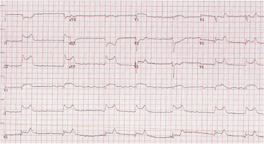
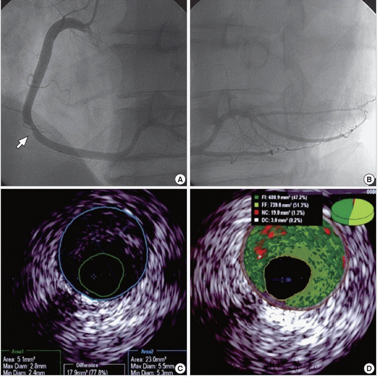
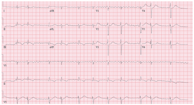
A 61-year-old male visited our emergency room presenting with substernal chest pain, which had developed one hour prior to presentation. He had been on medications for hypertension, hypertrophic cardiomyopathy and atrial fibrillation (Candesartan, 8 mg, Diltiazem, 90 mg, Carvedilol, 25 mg). An electrocardiogram (ECG) showed ST segment elevation in leads II and III, AVF and reciprocal ST depression in lead I, aVL and atrial fibrillation with a slow ventricular rhythm (Fig. 1). Cardiac enzyme (CK-MB: 1.95 ng/mL, troponin- I: 0.028 ng/mL) was mildly elevated. Two-dimensional echocardiogram revealed asymmetric left ventricular hypertrophy and akinesia in the right coronary artery (RCA) territories. After a temporary pacemaker was implanted, a coronary angiogram (CAG) showed thrombotic total occlusion in the proximal RCA without collateral flow. After wiring in the RCA, large amounts of thrombi were aspirated using Thrombuster (Kaneka Corp., Osaka, Japan). DC cardioversion was performed because ventricular fibrillation developed during PCI. Intracoronary abciximab, adenosine and nicorandil were injected, and the final CAG showed mild stenosis mid- RCA and thrombus remaining in the posterolateral branch (Fig. 2). Because CAG indicated TIMI III flow and poor patient status, we decided to initiate medical therapy in the coronary care unit. Seven days later, ECG demonstrated complete resolution of the elevated ST segment (Fig. 3) and follow-up CAG showed complete resolution of the thrombus of the posterolateral branch. Intravascular ultrasound (IVUS) showed moderate plaque in the middle RCA (minimal luminal area 5.1 mm2, plaque burden 77.8%) (Fig. 4). Therefore, we decided to maintain medical treatment without further interventions such as ballooning or stenting. The patient remained free of adverse cardiac events during a 2-month outpatient follow-up period.

Electrocardiography showed atrial fibrillation, ST elevation in leads II and III, AVF and reciprocal change in lead I, and AVL.

Thrombotic total occlusion in the proximal right coronary artery (RCA) (A). After wiring the RCA, thrombus aspiration was performed multiple times (B). Final coronary angiography showed mild stenosis in the middle RCA (C) and remaining thrombosis in the posterolateral branch (D).

Follow-up coronary angiogram showed no interval changes of mild stenosis in the middle right coronary artery (RCA) (A) and complete resolution of the thrombus in the posterolateral branch (B). Intravascular ultrasound showed moderate plaque in the middle RCA (minimal luminal area =5.1 mm2, plaque burden = 77.8%) (C, D).
Discussion
Ever since the publication of findings from the TAPAS trial, current guidelines have recommended thrombus aspiration as an effective treatment for patients with large thrombus burdens, followed by balloon angioplasty and stent implantation.[1,2] However, additional balloon angioplasty and stent implantation in culprit lesions involve additional risks of fragmentation of thrombi and embolization.[3] Distal embolization of atherothrombotic material is responsible for insufficient myocardial reperfusion after successful revascularization of the culprit lesion, and is associated with elevated mortality.[4]
Additional balloon angioplasty and stent implantation may not be helpful in selected patients,[5,6] such as patients with non-atherosclerotic causes for thrombus formation (embolism, malignancy, and acute endothelial dysfunction related to cocaine). In our case, atrial fibrillation might have been the cause of embolism. In addition, anti- platelet therapy is contraindicated in patients with high bleeding risk after balloon angioplasty or stent implantation. Additional ballooning or stent implantation involves procedural risks (bifurcation or ostial lesion). Finally, recent studies have shown that between 4% and 22% of culprit coronary lesions occur in non-significant obstructed plaques (<50%).[7] In such cases, it is difficult to determine the long-term risk/benefit ratio of additional ballooning or stent implantation after thrombus aspiration.
Although there have been no large scale randomized trials addressing this question, some studies and case reports suggest that thrombus aspiration can be safely used alone in selected patients.[5,6] Escaned et al.[6] performed PCI using thrombus aspiration alone in a sample of 28 patients with STEMI. During clinical follow-up (mean follow-up period: 40±23 months), thrombus aspiration alone was shown to be successful in all patients. There was one case of sudden cardiac death (4%) and three cases of non-cardiac death (11%). One patient (4%) was admitted with non-STEMI (new coronary angiogram without stenosis) and the remaining 22 (78.5%) remained asymptomatic and free of cardiac death. Kramer et al.[3] reported successful PCI using thrombus aspiration alone in a sample of 16 patients with STEMI (mean follow-up period: 1.3 years). Acceptable flow with minimal non-significant residual stenosis immediately after thrombus aspiration was observed in 14 patients (88%). In four patients (25%), repeat angiography was performed after several days and disappearance of the residual thrombus was confirmed in three of these patients. During follow-up, repeat target lesion revascularization was performed in one patient after 53 days. No recurrent myocardial infarctions were observed. Lesions favorable for aspiration only were found primarily in non-tortuous and non-calcified vessels with no residual lesions or mild residual stenosis (<40%), with smooth angiographic appearance of the vessel lumen and restoration of TIMI-3 flow.[8] Non-infarcted vessels that are angiographically normal confirm low total atherosclerotic burden. Therefore, thrombus aspiration alone could be safely performed as a primary revascularization procedure in selected STEMI patients.
In summary, we report a case of successful PCI with thrombus aspiration alone in a STEMI patient with chronic AF.
