Rapid Disappearance of Lupus Anticoagulant in COVID-19 Infection
Article information
Trans Abstract
Purpose
Severe acute respiratory syndrome-coronavirus 2 (SARS-CoV-2) infection affects various hematology-, hemostasis-, and thrombosis-related laboratory results, but controversies remain about its effect on lupus anticoagulant (LA). This study provides background content for LA in relation to coronavirus disease-2019 (COVID-19).
Methods
We retrospectively searched for patients with confirmed COVID-19 infections at Asan Medical Center from March 2020 to March 2021. Among the 151 confirmed patients, 74 who underwent LA testing were included in the study.
Results
Most patients had a mild infection course (64 survivors), except for 10 patients who died. LA was identified in 64.1% of survivors and in 30.0% of the patients who did not survive. The survivors’ LA confirmation tests were 95.12% positive for diluted Russell’s viper venom and 2.44% positive for silica clotting time. Of the 26 patients who underwent serial follow-up LA testing, nine (81.8%) showed a rapid disappearance of LA positivity within 12 weeks. There was only one case of LA persistence for > 12 weeks. There were no other antiphospholipid syndrome (APS)-related antibodies (anticardiolipin [ACA] antibodies or anti-beta-2-glycoprotein), except in one patient. The APS-related antibody ACA IgG was positive in only the one patient who demonstrated LA positivity for > 12 weeks.
Conclusion
A high proportion of LA positivity was found in patients with COVID-19. This is the first study to confirm that the proportion of LA positivity (78.4%) was high in Korean patients with COVID-19. Most LA positivity resolved within six weeks (81.8%), instead of by 12 weeks. The clinical significance of high LA-positive rates and rapid negative transitions in patients with COVID-19 needs further study.
Introduction
Coronavirus disease 2019 (COVID-19) is an infectious disease caused by a novel coronavirus (severe acute respiratory syndrome coronavirus 2 [SARS-CoV-2]). Severe hypercoagulable conditions and complex venous thrombosis are common [1-3]. Several recent studies have reported an elevated activated partial thromboplastin time (aPTT) in patients with COVID-19 with a thrombotic tendency, suggesting that the test results may be due to the presence of lupus anticoagulant (LA) [4-6]. In another study, patients severely ill with COVID-19 were identified as having antiphospholipid antibodies other than LA and concurrent coagulopathy [7]. Antiphospholipid antibodies, including LA, are associated with the diagnostic criteria for antiphospholipid syndrome (APS), which is characterized by a tendency toward thrombosis, and it is also important for ascertaining the meaning of LA in SARS-CoV-2 infection, which is characterized by thrombosis.
Here, we investigated the changes in LA and the characteristics of coagulation-related factors in patients with COVID-19 who underwent LA testing.
Methods
Study design
This study was conducted in patients diagnosed with COVID-19 from March 2020 to March 2021 at Asan Medical Center in Republic of Korea. It was performed as a retrospective chart analysis of patients with COVID-19 who had undergone LA testing. SARS-CoV-2 infection was confirmed in enrolled patients using real-time reverse-transcription polymerase chain reaction according to the World Health Organization’s interim guidance. Data on demographic information and coagulation test results were also obtained. The study was approved by the Ethics Committee of Asan Medical Center (S2021-0748-0001), and there are no conflicts of interest.
Methods of la testing and classification criteria
The LA assay used in this study followed the recommendations of the International Society for Thrombosis and Hemostasis [8]. Venous blood was collected in a 3.2% sodium citrate tube with an anticoagulant ratio of 9:1, and double centrifugation was performed within 5 h of blood collection to obtain platelet-poor plasma. The LA level was measured by an ACL TOP (Instrumentation Laboratory, MA, USA) using two tests with different principles: diluted Russell’s viper venom (DRVVT) and silica clotting time (SCT). DRVVT was considered the first test, and SCT, a test sensitive to aPTT, which is a silica activator, was considered the second test. The testing process was conducted in three stages: screening, mixing, and confirmation until LA positivity was confirmed.
Patients who were confirmed positive by LA testing were classified based on the period when they were confirmed to be negative. They were classified based on the APS diagnostic criteria for 12 weeks. If the test was performed after the scheduled follow-up date and a negative conversion was confirmed, it was classified as a “delayed F/U LA test” and placed in the group that transitioned to LA-negative within 12 weeks.
Statistical analysis
All comparisons of numeric parameters were conducted using the Mann-Whitney U test, and the results are shown as mean±standard deviation (SD). A P-value <0.05 was considered statistically significant. Data were analyzed using the Statistical Package for the Social Sciences, version 21.0.
Results
From March 2020 to March 2021, SARS-CoV-2 infection was identified in 151 patients; 74 of these underwent LA testing and were included in the analysis. The enrolled patients consisted of 64 survivors and 10 non-survivors with mean (±SD) ages of 57.9±18.1 years and 76.4±11.6 years, respectively. In survivors and non-survivors, 35 and 5 patients, respectively, were male. Since most enrolled patients had a mild course of infection, the analysis was performed primarily on the survivor group.
Of the survivors, 64.1% tested positive on the LA test, and most of them were positive for DRVVT. Only one patient showed LA positivity that persisted for ≥12 weeks, and most patients (96.2%) became LA-negative within 12 weeks (Fig. 1). In the survivors divided by LA results, additional analyses were performed, including coagulation parameters. A longer aPTT and higher C-reactive protein (CRP) and fibrinogen levels were identified in LA-positive survivors, and the results were statistically significant (Table 1).
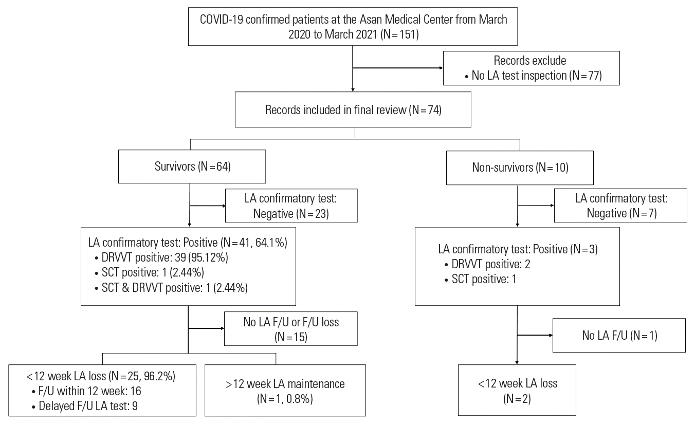
Enrollment and inclusion in the analysis.

Coagulation parameters of COVID-19 patients based on lupus anticoagulant testing
Changes in aPTT were examined in LA-positive patients. The analysis included 2 non-survivors and 16 survivors who successively underwent aPTT testing. When the change in aPTT was assessed, we confirmed that the aPTT tended to decrease as LA positivity changed to negativity (Fig. 2).

Decreasing aPTT trends following the conversion of positive lupus anticoagulant (LA) test results to negative in survivors (A) and non-survivors (B). The gray-shaded box represents LA-positive periods.
Discussion
LA can appear temporarily in viral infections because the infection acts as a trigger [4,9]. LA, which appears in some cases during infection, prolongs the aPTT. LA was also found in a high percentage of patients with COVID-19, and some had a risk of thrombosis [6]. In our study, 64.1% of patients with COVID-19 were LA-positive; these findings were consistent with those of previous studies. Interestingly, in our study, LA positivity was confirmed within 2 weeks of the first confirmation of COVID-19 infection (data not shown).
Therefore, it is vital to understand the importance of the presence of LA in patients with COVID-19. When COVID-19 patients experience severe thrombosis, prolongation of the aPTT due to LA positivity can cause problems with initiation of anticoagulant therapy. When using unfractionated heparin in patients with COVID-19, the aPTT can be prolonged due to the presence of LA, so clinicians must consider the “aPTT confounding” issue [6,10]. Bowles et al. [9] reported that most patients with COVID-19 with aPTT prolongation were positive for LA; thus, a prolonged aPTT should not interfere with anticoagulant therapy in the prevention and treatment of venous thrombosis in patients with COVID-19. In our study, prolongation of the aPTT was confirmed during the LA-positive period, and the aPTT normalized after LA disappearance.
However, it remains a point of debate whether the CRP, rather than LA, positively interferes with anticoagulation therapy, which can lead to aPTT prolongation; as a result, LA may be meaningless for patients with COVID-19 [5]. Due to the phospholipid binding affinity of CRP, the aPTT assay may show false prolongation, and the degree of increase may vary depending on the CRP level [11]. In our study, the range of CRP in the LA-negative group among the survivor groups was from 0.0–150 mg/L, while the LA-positive group ranged from 0.0–220 mg/L. However, the patients included in the analysis had mild infection, and the increase in CRP level was not significant. The aPTT was assessed in our study using the CS-5100 system (Siemens Healthcare Diagnostics, Erlangen, Germany), which uses ellagic acid as the activator. Another study reported that the same device manufacturer, which uses the same measurement principle, was not affected by an elevated CRP level [11,12]. Therefore, prolongation of aPTT should be considered as an effect of LA positivity instead of a result of CRP elevation.
Unlike the diagnostic criteria for APS, where antiphospholipid antibodies (including LA) need to be identified at intervals of ≥12 weeks, our analysis showed that 96.2% of the patients demonstrated a rapid disappearance of LA within 12 weeks. Only 1 patient was LA-positive for >12 weeks, and this patient was LA-positive 3 days before COVID-19 infection was confirmed. Thus, in all patients with COVID-19, LA resolved within 12 weeks, and 1 patient who remained LA positive for > 12 weeks may truly have had APS. Patients with COVID-19 show a tendency toward thrombosis, but LA resolves rapidly, so the relationship between LA and propensity for thrombosis is unclear. However, the significance of LA, which appears in a high proportion of patients with COVID-19, is unknown.
The limitations of our study are its small sample size and the small proportion of critically ill patients. More patients should be analyzed to determine the transient effects of LA observed in patients with COVID-19.
In conclusion, in SARS-CoV-2 infection, LA had a high rate of positivity within 2 weeks of infection confirmation and disappeared rapidly within 12 weeks, while LA positivity affected the aPTT results early during the infection. Therefore, care should be taken during the initial stages of patient management. Moreover, due to the rapid decline in LA, its correlation with thrombosis in patients with COVID-19 is unclear and requires further study.
Notes
The authors report no competing interests to declare.
Acknowledgements
The authors would like to thank all the enrolled patients for providing data for this study.